Cytotoxic constituents from Butea superba Roxb.
Nattaya Ngamrojanavanich a,∗, Aimon Loontaisong b, Somchai Pengpreecha a, Wichai Cherdshewasart c, Surachai Pornpakakul a, Khanitha Pudhom a, Sophon Roengsumran a, Amorn Petsom a
Research Centre for Bioorganic Chemistry, Faculty of Science, Chulalongkorn University, Pyathai Road, Pathumwan, Bangkok 10330, Thailand b Program of Biotechnology, Faculty of Science, Chulalongkorn University, Pyathai Road, Pathumwan, Bangkok 10330, Thailand
Department of Biology, Faculty of Science, Chulalongkorn University, Pyathai Road, Pathumwan, Bangkok 10330, Thailand
Received 20 January 2006; received in revised form 24 May 2006; accepted 20 July 2006
Available online 4 August 2006
Abstract
A carpin (3-hydroxy-9-methoxypterocarpan) (Medicarpin) (1) and four isoflavones, 7-hydroxy-4 -methoxy-isoflavone (Formononetin) (2); 7,4 – dimethoxyisoflavone (3); 5,4 -dihydroxy-7-methoxy-isoflavone (Prunetin) (4) and 7-hydroxy-6,4 -dimethoxyisoflavone (5) were isolated from the tuber roots of Butea Superba Roxb. Compounds 2 and 4 showed moderate cytotoxic activity on KB cell lines with IC50 (mM) values of 37.3 ± 2.5 and 71.1 ± 0.8 and on BC cell lines with IC50 (mM) values of 32.7 ± 1.5 and 47.3 ± 0.3, respectively.
© 2006 Elsevier Ireland Ltd. All rights reserved.
Keywords: Butea superba; Isoflavones; Carpin; Cytotoxicity
1. Introduction
With emphasis to the plants in the genus Butea, information of the isolated chemicals and/or bioactivities derived mostly from Butea frondosa Koen. ex Roxb. (synonym; Butea monosperma). The plant contained lectin in seeds and exhibited estrogenic and postcoital anti-conceptive activity in rats (Bhargava, 1986; Wongkham et al., 1994). Glucosides were also found in the plant extract (Gupta et al., 1970). The plant leaves exhibited anti-inflammatory effect (Mengi and Deshpande, 1999) and effect on stress, anxiety, and cognition in rats (Soman et al., 2004). The plant extract exhibited various bioactivities, including aphrodisiac activity in male rats (Ramachandran et al., 2004), protective role against thioacetamide-mediated hepatic alterations in Wistar rats (Sehrawat et al., 2006), in vivo anthelmintic activ-ity against Trichostrongylid nematodes in sheep (Iqbal et al., 2006), anti-diabetic in rats (Somani et al., 2006), anti-diarrheal activity in experimental animals (Gunakkunru et al., 2005), dermal wound healing in rats (Sumitra et al., 2005), anti-convulsive activity in laboratory animals (Kasture et al., 2000; Veena et al., 2002), anthelmintic activity (Prashanth et al., 2001) and anti-fungal (Bandara et al., 1989).
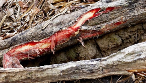
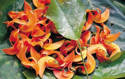
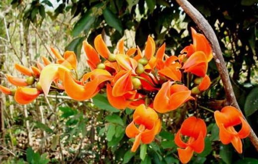
Butea superba Roxb. is a plant in the Family Leguminosae with the common name of “red Kwao Krua” in Thailand. The plant is abundantly distributed in the Thai deciduous forests in many parts of the country. This indigenous herb has been tradi-tionally consumed among Thai males for the purposes of rejuvenate as well as maintain sexual performance or prevent erectile dysfunction (Suntara, 1931). The Thai Butea superba Roxb. was found to contain flavonoid and flavonoid glycosides with potent cAMP phosphodiesterase inhibitory activity (Roengsumran et al., 2000). The plant crude extract exhibited significant inhibitory activity on acetylcholinesterase (Ingkaninan et al., 2003). The clinical trial of the plant powder as crude drug showed effective treatment of erectile dysfunction with a com-parable method with sildenafil treatment (Cherdshewasart and Nimsakul, 2003; Cappelleri and Rosen, 2005). Besides, Butea superba Roxb. exhibited anti-proliferation effect on the growth of MCF-7 and Hela cells in relation with a possible anti-estrogen mechanism or a potent cytotoxic effect (Cherdshewasart et al., 2004a,b). The Indian Butea superba Roxb. stems contained flavones glycoside and flavonol glycoside (Yadava and Reddy, 1998a,b). This suggests that the chemical constituents of this plant might partially show cytotoxic activity.
Therefore, this work describes the isolation and structural determination of the chemical constituents of this plant and their cytotoxic activity.
2. Materials and methods
2.1. General procedures
Melting points were determined on Electrothermal 9100. The optical rotations were determined on a Perkin-Elmer 341. Measurements of UV spectra were carried out on the Perkin-Elmer Lambda 25 UV–vis spectrophotometer. The 1H and 13C NMR spectra were recorded at 400.00 and 100.00 MHz, respectively, on a Varian Mercury-400 plus NMR spectrometer. MS spectra were measured with high-resolution electron spray/time of flight (HRES/TOF) mass spectrometer (Micro-mass UK Limited). Column chromatography was performed on silica gel 230–400 mesh for flash column chromatography (Isco CombiFlashTM GraduateTM).
2.2. Plant materials
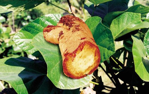
The tuber roots of Butea Superba Roxb. (Leguminosae) were collected from Lampang Province, Thailand in April 2002. The plant was authenticated by comparison with the herbarium col-lection number No. BCU 11046 in the Department of Botany, Faculty of Science, Chulalongkorn University, Bangkok, Thai-land.
2.3. Extraction and isolation
The dried tuber roots powder of Butea superba (5.5 kg) was extracted with MeOH (3× 15 L) and the methanolic extract (522 g) was re-extracted with hexane (2× 700 mL) and then CHCl3 (2× 700 mL). The hexane and CHCl3 extracts were evaporated under reduced pressure to give a crude hex-ane extract (1.2 g; 0.03%, w/w) and crude CHCl3 extract (40.0 g; 0.73%, w/w). The crude CHCl3 extract was separated by flash column chromatography and eluted with 100% of CH2Cl2, CH2Cl2–MeOH mixtures; the gradual concentration has increase in a stepwise of 1% MeOH. Compounds 1–5 (Fig. 1) were obtained from 1, 1.5 and 2% MeOH in CH2Cl2 and identified by comparison of their spectral data with reported values in the literatures.

2.4. Cell lines
KB, human epidermoid carcinoma of cavity (ATCC CCL-17); BC, breast cancer cell line; NCI-H 187, human small cell lung carcinoma (ATCC CRL-5804).
constituents from Butea superba
2.5. Cytotoxicity assay
KB and BC cell lines were determined by colorimetric cyto-toxicity assay that measured cell growth from cellular pro-tein content according to Skehan et al. (1990). Briefly, cells at a logarithimic growth phase were harvested and diluted to 105 cells/mL with fresh medium and gently mixed. Test com-pounds were dissolved with few drops of DMSO and diluted with water to make the concentration of DMSO less than 0.1%, added into 96-well plates in total volume 200 mL and incubated at 37 ◦C for 72 h. After incubation period, cells were fixed by 50% trichloroacetic acid, incubated at 4 ◦C for 30 min, washed with distilled water, dried at room temperature and stained with 0.05% (w/v) sulforhodamine B (SRB) dissolved in 1% acetic for 30 min. Unbound dye was removed with 1% acetic acid, and protein-bound dye was extracted with 10 mM Tris base [tris(hydroxymethyl)aminomethane] for determination of opti-cal density (OD) at 510 nm.
NCI-H 187 were determined by MTT (3-[4,5-dimethy-lhiazol-2-yl]-2,5-diphenyl tetrazolium bromide) assay previ-ously described in detail by Plumb et al. (1989). Briefly, cells were diluted to 105 cells/mL. Test compounds were dissolved with few drops of DMSO and diluted with water to make the con-centration of DMSO less than 0.1%, added into 96-well plates and incubated at 37 ◦C for 5 days. MTT solution (2 mg/mL) was added into each well and then incubated at 37 ◦C for 4 h. The MTT formazan crystals were dissolved in 200 mL for 100% DMSO and 25 mL of Sorensen’s glycine buffer. After 20 min, the optical density (OD) was measured with microtiter plate reader at wavelength of 510 nm.
Doxorubincin hydrochloride was used as a positive control and DMSO was used as a negative control.
2.6. Data analysis
Cell lines growth and growth inhibition were expressed in terms of mean (±1 of S.D.) absorbance units and/or percent-age of control absorbance (±1 of S.D.%) following subtraction of mean “background” absorbance. In addition, the IC50 was expressed as the sample concentration in micromolar that caused a 50% inhibition of growth compared with controls. The analysis was assisted with SPSS program.
3. Results and discussion
In the present study, the isolation of the chemical constituents with cytotoxicity from the tuber roots of Butea superba Roxb. were carried out. The dried tuber roots were extracted with methanol and re-extracted with hexane and chloroform at room temperature. The extracts were concentrated under reduce pres-sure, and then chloroform crude extract was chromatographed on flash column by eluting with gradients of dichloromethane and methanol to give compounds 1–5.
3-Hydroxy-9-methoxypterocarpan (Medicarpin) (1) (5 mg, 0.00009% yields from dried powder) was obtained from 1%
MeOH in CH2C12 as white solid mp 127–129 ◦C, UV(CHC13) 236.06 and 265.93 nm (log ε 4.14 and 4.34) at 27 ◦C; [α]D 265
−223.60 (c 0.01 g/100 mL, CH2Cl2). The structure of compound 1 was also confirmed by 2D-NMR, which is in agreement to those previously reported (Stadler et al., 1994; Chan et al., 1998; Chang et al., 1997; Konoshima et al., 1997).
7-Hydroxy-4 -methoxy-isoflavone (Formononetin) (2) (6.0 mg, 0.00013% yields from dried powder) was obtained from 2% MeOH in CH2Cl2 as orange solid mp 261–262 ◦C, UV(CHC13) λmax 273.00 and 295.01 nm (log ε 3.19 and 3.34) at 27 ◦C; [α]D265 −32.85 (c 0.01 g/100 mL, CHCl3). The structure of compound 2 was also confirmed by 2D-NMR, which is in agreement to those previously reported (Lopes et al., 1999; Khan et al., 2000).
7,4 -Dimethoxyisoflavone (3) (1.2 mg, 0.000009% yields from dried powder) was obtained from 1.5% MeOH in CH2C12
as yellow solid mp 162–163 ◦C, UV(CHC13) λmax 239.05 and 300.97 nm (log ε 5.24 and 5.20) at 27 ◦C; [α]D265 −19.68 (c
0.05 g/100 mL, CHCl3). The structure of compound 3 was also confirmed by 2D-NMR, which is in agreement to those previ-ously reported (Ingham et al., 1981; Veitch et al., 2003).
5,4 -Dihydroxy-7-methoxy-isoflavone (Prunetin) (4) (6.0 mg, 0.00013% yields from dried powder) was obtained from 1% MeOH in CH2C12 as brown solid mp 239–240 ◦C, UV (CHCl3) λmax 287.94 nm (log ε 3.21) at 27 ◦C; [α]D265 −19.90 (c 0.04 g/100 mL, CHCl3). The structure of compound 4 was also confirmed by 2D-NMR, which is in agreement to those previously reported (Baker et al., 1953; Tanaka et al.,1998; Talukdar et al., 2000).
7-Hydroxy-6,4 -dimethoxyisoflavone (5) (4.0 mg, 0.00007% yields from dried powder) was obtained from 1.5% MeOH in CH2C12 as yellow solid mp 228–229 ◦C, UV(CHC13) λmax 240.93, 270.97 and 315.07 nm (log ε 5.04, 4.99 and 5.34) at 27 ◦C; [α]D265 −5.65 (c 0.08 g/100 mL, CHCl3). The structure of compound 5 was also confirmed by 2D-NMR, which is in agreement to those previously reported (McMurry and Theng,1960; Herath et al., 1998).
The cytotoxicity of compounds 1–5 was assessed by SRB (for KB and BC cell lines) and MTT assay (for NCI-H187 cell line). Compounds 2 and 4 showed moderate cytotoxic activ-ity on KB cell lines with IC50 (mM) values of 37.3 ± 2.5 and 71.1 ± 0.8 and on BC cell lines with IC50 (mM) values of 32.7 ± 1.5 and 47.3 ± 0.3, respectively. Compounds 1, 3 and 5 were considered to be inactive (IC50 > 100 mM). Under the same test condition, doxorubicin hydrochloride exhibited cyto-
toxic activity against KB cell lines, BC cell lines and NCI-H187 cell line at IC50 (mM) values of 0.3, 0.4 and 0.5, respectively (Table 1).

Compounds 1–5 have been reported in many plant species and they exhibit various biological activities. Compound 1 showed anti-microbial activity (Stadler et al., 1994), anti-tumor promot-ing activity (Konoshima et al., 1997) and apoptosis-inducing effects on human PBMCs (Zhen-Li et al., 2002). Compound 2 showed anti-fungal activity against Cladosporium cladosporioides at 10-fold higher than the positive control Nystatin (Lopes et al., 1999). Compound 3 showed anti-giardial activity (Khan et al., 2000) and anti-plasmodial activity against Plasmodium falciparum (Kraft et al., 2001). Compound 4 showed only moderate anti-plasmodial activity against Plasmodium fal-ciparum (Carola et al., 2000). Therefore, this work reveals the chemical constituents and their moderate to weak cytotoxic-ity of Butea superba Roxb. for the first time. Moreover, other biological activities could be realized from constituents from Butea superba.
Acknowledgements
We thank the Higher Education Commission Grants for Graduate Dissertation, Chulalongkorn University. The Rachadapiseksompoj Endowment, Chulalongkorn University are also gratefully acknowledged.
References
Baker, W., Chadderton, J., Harborne, J.B., Ollis, W.D., 1953. A new synthesis of isoflavones. Part I. Journal of Chemical Society, 1852–1860.
Bandara, B.M., Kumar, N.S., Samaranayake, K.M., 1989. An antifungal con-stituent from the stem bark of Butea monosperma. Journal of Ethnopharma-cology 25, 73–75.
Bhargava, S.K., 1986. Estrogenic and postcoital anticonceptive activity in rats of butin isolated from Butea monosperma seed. Journal of Ethnopharmacology 18, 95–101.
Cappelleri, J.C., Rosen, R.C., 2005. The sexual health inventory for men (SHIM): a 5-year review of research and clinical experience. International Journal of Impotence Research 17, 207–319.
Carola, K., Kristina, J., Karsten, S., Mahabir, P.G., Ulrich, B., Eckart, E., 2000. Antiplasmodial activity of isoflavones from Andira inermis. Journal of Ethnopharmacology 73, 131–135.
Chan, S.C., Chang, Y.S., Wang, J.P., Chen, S.C., Kuo, S.C., 1998. Three new flavonoids and antiallergic, anti-inflammatory constituents from the heart-wood of Dalbergia odorifera. Planta Medica 64, 153–158.
Chang, L.C., Gerhauser, C., Song, L., Farnsworth, N., Pezzuto, J.M., Kinghorn, A.D., 1997. Activity-guided isolation of constituents of Tephrosia purpurea with the potential to induce the phase II enzyme, quinone reductase. Journal of Natural Products 60, 869–873.
Cherdshewasart, W., Cheewasopit, W., Picha, P., 2004a. The differential anti-proliferation effect of white (Pueraria mirifica), red (Butea superba), and black (Mucuna collettii) Kwao Krua plants on the growth of MCF-7 cells. Journal of Ethnopharmacology 93, 255–260.
Cherdshewasart, W., Cheewasopit, W., Picha, P., 2004b. Anti-proliferation effects of the white (Pueraria mirifica), red (Butea superba) and black (Mucuna collettii) Kwao Krua plants on the growth of HeLa cells. Journal of Scientific Research (Chulalongkorn University) 29, 27–32.
Cherdshewasart, W., Nimsakul, N., 2003. Clinical trial of Butea superba, an alternative herbal treatment for erectile dysfunction. Asian Journal of Andrology 5, 243–246.
Gunakkunru, A., Padmanaban, K., Thirumal, P., Pritila, J., Parimala, G., Ven-gatesan, N., Gnanasekar, N., Perianayagam, J.B., Sharma, S.K., Pillai, K.K., 2005. Anti-diarrhoeal activity of Butea monosperma in experimental ani-mals. Journal of Ethnopharmacology 98, 241–244.
Gupta, S.R., Ravindranath, B., Seshadri, T.R., 1970. The glucosides of Butea monosperma. Phytochemistry 9, 2231–2235.
Herath, H.M.T.B., Dassanayake, R.S., Priyadarshani, A.M.A., De Silva, S., Wannigama, G.P., Jamie, J., 1998. Isoflavonoids and a pterocarpan from Gliricidia sepium. Phytochemistry 47, 117–119.
Ingham, J.L., Keen, N.T., Mulheirn, L.J., Lyne, R.L., 1981. Inducibly formed isoflavonoids from leaves of soybean. Phytochemistry 20, 795–798.
Ingkaninan, K., Temkitthawon, P., Chuendhom, K., Yuyaem, T., Thongnoi, W., 2003. Screening for acetylcholinesterase inhibitory activity in plants used in Thai traditional rejuvenating and neurotic remedies. Journal of Ethnophar-macology 89, 261–264.
Iqbal, Z., Lateef, M., Jabbar, A., Ghayur, M.N., Gilani, A.H., 2006. In vivo anthelmintic activity of Butea monosperma against Trichostrongylid nema-todes in sheep. Fitoterapia 77, 137–140.
Kasture, V.S., Chopde, C.T., Deshmukh, V.K., 2000. Anticonvulsive activity of Albizzia lebbeck, Hibiscus rosasinesis and Butea monosperma in experimen-tal animals. Journal of Ethnopharmacology 71, 65–75.
Khan, I.A., Avery, M.A., Burandt, C.L., Goins, D.K., Mikell, J.R., Nash, T.E., Azadegan, A., Walker, L.A., 2000. Antigiardial activity of isoflavones from Dalbergia frutescens bark. Journal of Natural Products 63, 1414– 1416.
Konoshima, T., Takasaki, M., Kozuka, M., Tokuda, H., Nishino, H., Mat-suda, E., Nagai, M., 1997. Anti-tumor promoting activities of isoflavonoids from Wistaria brachybotrys. Biological Pharmaceutical Bulletin 20, 865–868.
Kraft, C., Jenett-Siems, K., Siems, K., Solis, P.N., Gupta, M.P., Bienzle, U., Eich, E., 2001. Andinermals A–C, antiplasmodial constituents from Andira inermis. Phytochemistry 58, 769–774.
Lopes, N.P., Kato, M.J., Yoshida, M., 1999. Antifungal constituents from roots of Virola surinamensis. Phytochemistry 51, 29–33.
McMurry, T.B.H., Theng, C.Y., 1960. The constitution and synthesis of afro-mosin. Journal of Chemical Society 12, 1491–1498.
Mengi, S.A., Deshpande, S.G., 1999. Anti-inflammatory activity of Butea fron-dosa leaves. Fitoterapia 70, 521–522.
Plumb, J.A., Milroy, R., Kaye, S.B., 1989. Effects of the pH depen-dence of 3-(4,5-dimethylthiazol-2-yl)-2,5-diphenyl-tetrazolium bromide-formazan absorption on chemosensitivity determined by a novel tetrazolium-based assay. Cancer Research 49, 4435–4440.
Prashanth, D., Asha, M.K., Amit, A., Padmaja, R., 2001. Anthelmintic activity of Butea monosperma. Fitoterapia 72, 421–422.
Ramachandran, S., Sridhar, Y., Kishore Gnana Sam, S., Saravanan, M., Thomas Leonard, J., Anbalagan, N., Sridhar, S.K., 2004. Aphrodisiac activity of Butea frondosa Koen. ex Roxb. extract in male rats. Phytomedicine 11, 165–168.
Roengsumran, S., Petsom, A., Ngamrojanavanich, N., Rugsilp, T., Sit-tiwicheanwong, P., Khorphueng, P., Cherdshewasart, W., Chaichan-tipyuth, C., 2000. Flavonoid and flavonoid glycoside from Butea superba Roxb. and their cAMP phosphodiesterase inhibitory activity. Journal of Scientific Research (Chulalongkorn University) 25, 169– 176.
Sehrawat, A., Khan, T.H., Prasad, L., Sultana, S., 2006. Butea monosperma and chemomodulation: Protective role against thioacetamide-mediated hepatic alterations in Wistar rats. Phytomedicine 13, 157–163.
Skehan, P., Storeng, R., Scudiero, D., Monks, A., McMahon, J., Vistica, D., Warren, J.T., Bokesch, H., Kenney, S., Boyd, M.R., 1990. New colorimetric cytotoxicity assay for anticancer drug screening. Journal of the National Cancer Institute 82, 1107–1112.
Soman, I., Mengi, S.A., Kasture, S.B., 2004. Effect of leaves of Butea frondosa on stress, anxiety, and cognition in rats. Pharmacology Biochemistry and Behavior 79, 11–16.
Somani, R., Kasture, S., Singhai, A.K., 2006. Antidiabetic potential of Butea monosperma in rats. Fitoterapia 77, 86–90.
Stadler, M., Dance, E., Anke, H., 1994. Nematicidal activities of two phytoalexins from Tavernaria abyssinica. Planta Medica 60, 550– 552.
Sumitra, M., Manikandan, P., Suguna, L., 2005. Efficacy of Butea monosperma on dermal wound healing in rats. The International Journal of Biochemistry and Cell Biology 37, 566–573.
Suntara, A., 1931. The Remedy Pamphlet of Kwao Krua Tuber of Luang Anusarnsuntarakromkarnpiset, Chiang Mai. Upatipongsa Press, Chiang Mai, Thailand.
Talukdar, A.C., Jain, N., De, S., Krishnamurty, H.G., 2000. An isoflavone from Myristica malabarica. Phytochemistry 53, 155–157.
Tanaka, T., Ohyama, M., Iinuma, M., Shirataki, Y., Komatsu, M., Burandt, C.L., 1998. Isoflavonoids from Sophora secundiflora, S. arizonica and S. gyp-sophila. Phytochemistry 48, 1187–1193.
Veena, S., Kasture, S.B., Chopde, C.T., 2002. Anticonvulsive activity of Butea monosperma flowers in laboratory animals. Pharmacology Biochemistry and Behavior 72, 965–972.
Veitch, N.C., Sutton, P.S.E., Kite, G.C., Irelandt, H.L., 2003. Six new isoflavones and a 5-deoxyflavonol glycoside from the leaves of Ateleia herbertsmithii. Journal of Natural Products 66, 210–216.
Wongkham, S., Wongkham, C., Trisonthi, C., Boonsiri, P., Simasathiansophon, S., Atisook, K., 1994. Isolation and properties of a lectin from the seeds of Butea monosperma. Plant Science 103, 121–126.
Yadava, R.N., Reddy, K.I., 1998a. A novel glycoside from the stem of Butea superba. Fitoterapia I. 19, 269–270.
Yadava, R.N., Reddy, K.I., 1998b. A new bio-active flavonol glycoside from the stems of Butea superba Roxb. Journal of Asian Natural Products Research 1, 139–145.
Zhen-Li, L., Tanaka, S., Horigome, H., Hirano, T., Oka, K., 2002. Induction of apoptosis in human lung fibroblasts and peripheral lymphocytes in vitro by shosaiko-to derived phenolic metabolites. Biological Pharmaceutical Bul-letin 25, 37–41.
constituents from Butea superba
constituents from Butea superba
constituents from Butea superba

